Science
The Menicon BCLA 2025 Showcase: orthokeratology, ocular health and efficacy

Sponsored by
In this article:
This article provides a summary of the Menicon poster presentations, highlighting key findings and their implications for clinical practice in myopia management and orthokeratology.
- BEST POSTER WINNER: Allergy Effects in Ortho-k Wearers
- 1ST RUNNER UP BEST POSTER: Corneal Endothelium After 36 Months of Ortho-k
- HOT TOPIC AWARD: Oral-resident bacteria and Acanthamoeba Risk in Ortho-k Cases
- Cleaning Efficacy of Ortho-k Care Solutions
- Peripheral Refraction: Ortho-k vs Myopia Control Spectacles
- Ortho-k Effects on Axial Length and Myopia Progression
- Eleven years of Ortho-k for Childhood Myopia Control
- Key takeaways
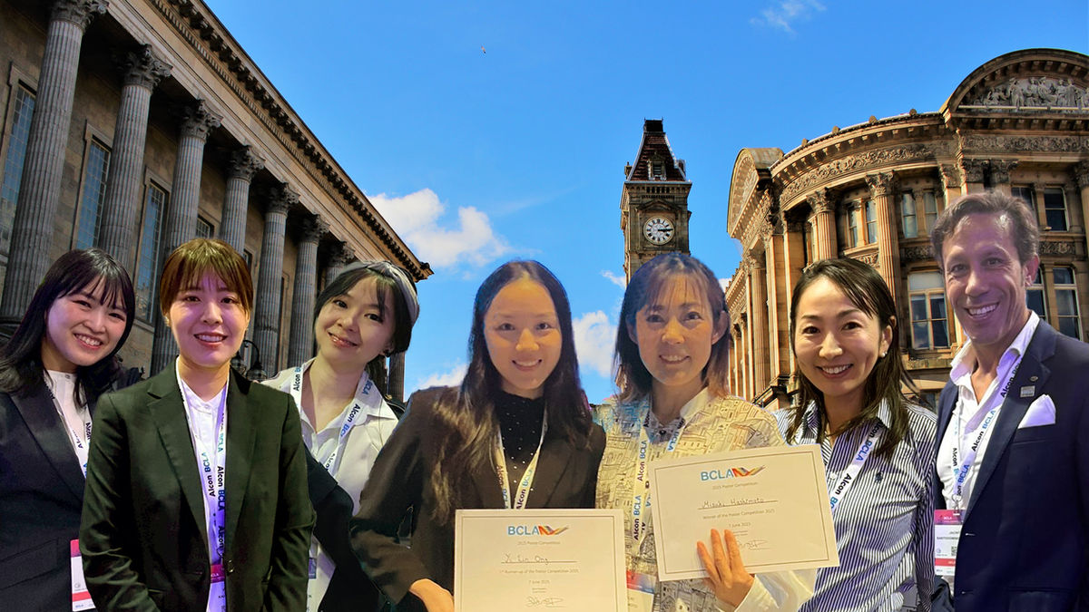
The British Contact Lens Association (BCLA) Clinical Conference and Exhibition was held in Birmingham, UK from 5–7 June 2025. This biennial event is a key platform for presenting advancements in contact lens research, clinical practice, and ocular health. Menicon contributed significantly to the scientific program, presenting seven abstracts focused on orthokeratology and ocular health. Their research was recognised with multiple awards, including Best Poster, First Runner-Up Poster, and a Hot Topic selection—reflecting both the breadth and clinical relevance of their work.
BEST POSTER WINNER: Clinical Observations and Lens Assessments in Orthokeratology Lens Wearers with Allergic Symptoms
Authors: Mizuki Hashimoto (pictured left)1,2, Saiko Matsumura (pictured right)1, Takashi Itokawa1, Kazuhiko Dannoue3, Satoshi Ohtsu3, Kazuma Niimura3, Madoka Yoshimitsu2, Keiji Sugimoto2, Yuichi Hori1
Affiliations:
- Department of Ophthalmology, Toho University Faculty of Medicine, Tokyo, Japan
- Global R&D, Menicon Co., Ltd., Aichi, Japan
- Dannoue Eye Clinic, Kanagawa, Japan
Summary
Orthokeratology (ortho-k) is an effective strategy for myopia control, but allergic symptoms in some wearers can compromise comfort, compliance, and long-term success. This case series reviewed eight pediatric ortho-k wearers who had been using ortho-k for an average of 413.25 days and had developed allergic symptoms, aiming to explore associated lens findings and care behaviours. All patients showed signs of allergic conjunctivitis, and most also had corneal epithelial damage. In three cases, symptoms led to discontinuation. Lens deposits were evaluated using imaging and quantified with software, revealing greater cloudiness in lenses worn for over a year or without protein removal. Some patients also experienced reduced visual acuity from induced astigmatism as a result of lens decentration. Findings suggest that lens material, wear duration, and compliance with lens care influence the risk of allergic symptoms and deposit formation. Regular monitoring, timely lens replacement, and reinforcing care routines are essential to maintain ocular health and minimise complications.
Abstract
Purpose: Orthokeratology (Ortho-K) lenses are effective for myopia control. However, some wearers may experience allergic reactions, leading to discontinuation. Clinical findings and observations of lens deposits related to Ortho-K lens wearers with allergic symptoms are not well understood.
The purpose was to report clinical observations, including slit-lamp findings, visual acuity, and patient-reported symptoms, along with lens assessments in Ortho-K lens wearers presenting with allergic symptoms.
Methods: Clinical observations, lens wear duration, and care adherence were reviewed in 8 Ortho-K lens wearers who presented allergic symptoms. Cases included discontinuation due to chronic allergies. Images to observe lens condition were obtained by stereomicroscopy and deposits were visually inspected. Deposits were quantified using the Volume/Pixel (V/P) value representing cloudiness per pixel to calculate the total cloudiness volume, derived from image analysis software called JustTLC.
Results: Eight cases (3 males, 5 females) were reported, with an average age of 10.88 years and an average lens wear duration of 413.25 days. All subjects exhibited some allergic symptoms, with three cases leading to discontinuation. Allergic conjunctivitis was observed in all cases, with 7 also showing corneal epithelial damage. Lens displacement occurred in 2 patients, leading to induced astigmatism, and decreased visual acuity. The average V/P value was 0.051±0.023, with higher values in lenses worn for over a year. V/P value varies depending on the type of lens worn, lens wear duration, and cases without protein removal had higher V/P values.
Conclusion: It had been indicated that contamination of Ortho-K lenses in wearers with allergic symptoms was correlated with the type of lens worn, lens wear duration, and lens care behavior. An appropriate lens exchange period and proper lens care, along with providing active guidance to improve patient compliance, are crucial. If these measures do not resolve the issue, discontinuation may also need to be considered.
Figure: Average Volume/Pixel (V/P) values—cloudiness per pixel from stereomicroscopy images —was used as an index of ortho-k lens deposit load across wear and care factors. Higher V/P (more cloudiness) occurred with above-average wearing periods, lens brand B, and omission of protein-removal steps, compared with shorter wear, lens brand A, and regular protein removal.
1ST RUNNER UP FOR BEST POSTER: Morphology of Corneal Endothelial Cells in Myopic Children in Kuala Lumpur after 36 months of wearing Orthokeratology Lenses
Authors: Yi Lin Ong (pictured)1, Bariah Mohd-Ali1, Yu Chen Low1
- Optometry and Vision Science Program, Centre for Community Health Studies (ReaCH), Faculty of Health Sciences, Universiti Kebangsaan Malaysia
Summary
Evaluating long-term ocular safety is essential in managing pediatric patients using ortho-k. This prospective study followed 20 myopic children aged 6–12 years over a 36-month period of ortho-k wear (Menicon Z lenses), assessing anterior segment health and myopia progression. Significant improvements in unaided visual acuity and a reduction in spherical equivalent refraction (SER) were observed after 12 months, with a consistent flattening of corneal curvature and slowed axial elongation over the full study period. Axial length increased by an average of 0.22 mm over three years in total, after being stable in the first year, indicating an annual progression rate of 0.11 mm in the subsequent two years. Importantly, corneal endothelial cell density, hexagonal cell percentage, and coefficient of variation remained stable throughout, suggesting no significant morphological changes. Minor complications such as corneal staining and infections were noted but were not statistically or clinically significant. Overall, these findings support that long-term ortho-k wear is a safe option for myopic children, with no permanent changes to corneal structure and stable ocular health parameters over time—reassuring clinicians and families of its long-term safety in myopia management.
Abstract
Purpose: The study aimed to evaluate the long-term effects of orthokeratology (Ortho-K) lenses on corneal endothelial morphology and overall anterior segment health in myopic children over a 36-month period, while monitoring for potential complications.
Methods: A total of 20 myopic children aged 6–12 years, who had been wearing Ortho-K lenses (Menicon Z) for 36 months were included. Assessments were conducted at baseline, 12 months, and 36 months including endothelial cell density (ECD), hexagonal cell percentage (HEX), coefficient of variation (COV) and central corneal thickness (CCT). Additional parameters such as Unaided VA, Spherical Equivalent (SE), axial length (AL) and corneal curvature (FK) were analyzed. The incidence of complications was also recorded.
Results: After 12 months of Ortho-K wear, unaided visual acuity improved significantly, accompanied by a reduction in SE (p < 0.05). From 12 to 36 months, SE showed a slight increase of -0.16±0.06D (p < 0.05), while corneal curvature continued to flatten significantly (p < 0.001). AL initially decreased at 12 months but increased by 0.22±0.004mm at 36 months (p < 0.001), with a progression rate of 0.11mm/year, indicating slowed myopia progression. Corneal endothelial morphology remained stable throughout the study, with no significant changes in ECD, COV, or HEX. Mild complications, including corneal staining and infections, were observed but were not statistically significant.
Conclusion: Ortho-K effectively slowed myopia progression over 36 months without significant adverse effects on corneal endothelial morphology. While minor complications occurred, they were not clinically significant. These findings support the long-term safety of Ortho-K in myopic children.
HOT TOPIC AWARD: Effect of Oral-resident Bacterial Coexistence on Biofilm-forming Potency of Staphylococcus epidermidis Found on Used Orthokeratology Lens Cases and Acanthamoeba castellanii Proliferation
Authors: Ai Watanabe (pictured)1, Yuna Kimura1, Taizo Sumide1
- Global R&D, Menicon Co., Ltd., Aichi, Japan
Summary
Understanding environmental and microbial factors that contribute to lens case contamination is critical in preventing ortho-k–related microbial keratitis. This in vitro study investigated whether the presence of oral-resident bacteria influences the biofilm-forming ability of Staphylococcus epidermidis—isolated from used ortho-k lens cases—and the proliferation of Acanthamoeba castellanii. Three oral-resident species (Streptococcus salivarius, S. parasanguinis, and S. oralis) were co-cultured with S. epidermidis or A. castellanii in separate experiments. Biofilm formation was significantly increased when S. epidermidis coexisted with S. salivarius or S. oralis. Additionally, A. castellanii proliferation was significantly enhanced in the presence of all three bacterial species at higher concentrations (≥10⁷ CFU/mL). This study highlights an under-recognised risk factor in ortho-k lens care: contamination from oral-resident bacteria and suggests that everyday hygiene habits—such as storing lens cases near bathroom sinks where oral bacteria may be dispersed—can potentially increase the risk of microbial keratitis. Simple changes in storage location and hygiene routines may reduce the risk of ortho-k-related microbial complications.
Abstract
Purpose: To evaluate how oral-resident bacterial coexistence could affect biofilm-forming potency of Staphylococcus epidermidis isolated from used orthokeratology lens cases and Acanthamoeba castellanii proliferation.
Methods: Three kinds of oral-resident bacteria (Streptococcus salivarius and Streptococcus parasanguinis isolated from used orthokeratology lens cases, and Streptococcus oralis ATCC 35037) were selected. In experiment 1, each bacterium in a concentration of approximately 101 to 104 CFU/mL coexisted with S. epidermidis isolated from used orthokeratology lens cases through cell culture inserts for 2 days. Subsequently, biofilm-forming potency of S. epidermidis was quantified using the crystal violet assay. In experiment 2, each bacterium in a concentration of approximately 102 to 109 CFU/mL was inoculated with A. castellanii ATCC 50370 in a concentration of 5×10³ CFU/mL. After incubation for 7 days, A. castellanii proliferation was quantified by counting the cell number. In both experiments, no coexistence of oral-resident bacteria was used as control.
Results: In experiment 1, biofilm-forming potency of S. epidermidis was enhanced under coexistence with S. salivarius and S. oralis, respectively (all p≤0.05, Dunnett test, vs control). In experiment 2, A. castellanii proliferation was increased under coexistence with all three oral-resident bacteria in a concentration of 107 CFU/mL or more, respectively (all p≤0.01, Dunnett test, vs control).
Conclusions: This study showed that oral-resident bacterial coexistence could enhance biofilm-forming potency of S. epidermidis isolated from used orthokeratology lens cases and A. castellanii proliferation. Storage of contact lens cases around bathroom washstands could increase the risk of contamination with oral-resident bacteria. As such, contact lens cases should be managed in a hygienic manner including the proper storage location to prevent contamination with microorganisms, especially oral-resident bacteria. In addition, it could contribute to reducing the risk of orthokeratology-related microbial keratitis.
Cleaning efficacy and anti-bacterial properties of contact lens care solutions for orthokeratology lenses
Authors: Yui Kahara (pictured)1, Sayaka Goto1, Taizo Sumide1
- R&D Center, Menicon Co. Ltd., Kasugai, Aichi, Japan
Summary
This in vitro study evaluated the cleaning effectiveness and anti-adhesion properties of various contact lens care solutions on ortho-k lenses exposed to tear film–like deposits. Lenses were divided into the following groups:
- (A): No cleaning (control)
- (B1): Multipurpose solution 1 with rubbing — reduced deposits to 8.9% and P. aeruginosa adhesion to 14.0%
- (B2): Multipurpose solution 2 with rubbing — reduced deposits to 4.7% and adhesion to 4.5%
- (B3): Hydrogen peroxide with rubbing — reduced deposits to 5.3%, but adhesion remained relatively high at 86.0%
- (B4): Chlorine-based solution without rubbing — reduced deposits to 3.9% and adhesion to 41.8%
- (C): Unworn lenses — baseline levels of 2.3% deposits and 28.4% adhesion
All treatment groups showed a reduction in surface deposits and Pseudomonas aeruginosa adhesion compared to the untreated control. The best overall performance was observed with multipurpose solutions that included a rubbing step, particularly (B2). The findings support that daily rubbing and periodic chlorine-based disinfection are effective strategies for maintaining ortho-k lens hygiene and minimizing bacterial adhesion—important steps in reducing the risk of microbial keratitis in ortho-k wearers.
Abstract
Purpose: To evaluate the cleaning efficacy and anti-adhesion properties against Pseudomonas aeruginosa of contact lens care solutions on orthokeratology (orthoK) lenses in vitro.
Methods: Deposited orthoK lenses were prepared by soaking in an artificial solution mimicking that of the tear film for 7hr at 80℃ and randomly allocated to three groups, including a group that acted as a control in which lenses were not cleaned (A); a second group in which lenses were treated with contact lens cleaning/disinfecting solutions (two types of multi-purpose solution including a rubbing step: (B1) and (B2); hydrogen peroxide solution including a rubbing step: (B3); chlorine-based solution without rubbing step: (B4)); and a third group in which lenses were unworn orthoK lenses (C). After lenses treatment with each care solution according to instructions for use, in experiment 1, the deposits onto each orthoK lens were quantified as the total volume of cloudiness over the lens surface in terms of volume per unit pixel (V/P value) using image analysis (n=4). In experiment 2, each orthoK lens was inoculated with Pseudomonas aeruginosa for 4hr at 37℃ and the number of Pseudomonas aeruginosa adhesion onto the lens surface was quantified by reverse transcriptase-polymerase chain reaction (n=3).
Results: Compared with (A), the V/P value and the adhesiveness of Pseudomonas aeruginosa for the different orthoK lens conditions reduced to 8.9% and 14.0%, respectively in (B1); to 4.7% and 4.5%, respectively in (B2); to 5.3% and 86.0%, respectively in (B3); to 3.9% and 41.8%, respectively in (B4); and to 2.3% and 28.4%, respectively in (C).
Conclusions: Daily rubbing of orthoK lenses prior to using a cleaning/disinfecting solution and the regular use of chlorine-based solution were effective in removing deposits and keeping lenses clean, thus minimizing the adhesion of Pseudomonas aeruginosa onto the lens surface.
Comparison of peripheral refraction between orthokeratology and myopia control spectacle lens wearers at 12 months
Authors: Bariah Mohd-Ali1, Low Yu Chen1, Syarifah Faiza Syed Mohd Dardin1, Mizhanim Mohamad Shahimin1
- Optometry and Vision Science Program, Centre for Community Health Studies (ReaCH),
Faculty of Health Science, Universiti Kebangsaan Malaysia
Summary
Peripheral retinal profile may influence treatment outcomes in myopia control, yet different interventions can produce varying effects on retinal shape. This retrospective study compared the impact of ortho-k and Defocus Incorporated Multiple Segments (DIMS) spectacle lenses on peripheral refraction (PR) over 12 months in 65 myopic children aged 8–10 years. PR was measured across the horizontal meridian under cycloplegia. Both groups showed comparable myopia control efficacy (~63% reduction in progression), but ortho-k wearers demonstrated significantly greater changes in peripheral refraction across all measured eccentricities compared to DIMS wearers (p<0.001). These findings suggest that ortho-k induces a more prolate retinal shape change, while DIMS wear results in a comparatively oblate profile. Although both interventions were similarly effective in slowing axial progression, they appear to exert their optical effects through different peripheral mechanisms.
Abstract
Purpose: To compare the impact of wearing orthokeratology (Ortho-K) and Defocus Incorporated Multiple segments (DIMS) spectacle lens for 12 months on peripheral refraction (PR) in myopic children.
Methods: This retrospective study using clinical records from children attending the university’s Optometry Clinic in Kuala Lumpur, involving 65 children (45 Ortho-K, 20 DIMS). Participants were children aged 8-10 years, with refractive error of -0.50 to -5.00 D and were included after 12 month completion of follow-up. Cycloplegic PR was conducted using autorefractometer along horizontal meridian at 10° intervals up to 30° nasal (N) and temporal (T). The change in refraction was compared using t-tests
Results: At 12 months, both lenses successfully controlled myopia progression (62.5% OK, 63.5% DIMS). Significant difference was noted in the PR at all eccentricities between both groups (p<0.001), with higher change in the Ortho-K than DIMS wearers. This indicates a more oblate posterior retinal shape in DIMS than Ortho-K wearers after 12 months of wearing interventions.
Conclusions: Both OK and DIMS lenses controlled myopia progression at a similar rate, but with different impact on PR.
The Impact of Orthokeratology on Peripheral Refraction, Axial Length and myopia progression rate in Chinese Myopic Children: A 36-Month Study
Authors: Yu Chen Low (pictured)1, Bariah Mohd-Ali1, Mizhanim Mohamad Shahimin1
- Optometry and Vision Science Program, Centre for Community Health Studies (ReaCH),
Faculty of Health Science, Universiti Kebangsaan Malaysia
Summary
Long-term evaluation of peripheral optical changes is important to understand the sustained effects of orthokeratology on myopia control. This 36-month prospective study assessed peripheral refraction (PR), axial length, and myopia progression in 25 myopic children fitted with Ortho-K lenses. Measurements were taken centrally and across horizontal eccentricities (±10°, 20°, 30°) using cycloplegic autorefraction and A-scan biometry. Central refraction improved significantly over time, with a low progression rate of –0.08D/year. Axial elongation was significantly reduced, averaging 0.11 mm/year. PR shifted towards more myopic defocus across all eccentricities at 12 months. From 12 to 36 months, a further significant myopic shift was observed nasally, but not temporally. This study adds to the evidence that ortho-k can sustainably reduce axial elongation while maintaining or enhancing peripheral myopic defocus, especially nasally.
Abstract
Purpose: This is a continuation study aimed to evaluate the effects of orthokeratology (Ortho-K) lenses on peripheral refraction (PR), axial length (AL) elongation and myopia progression rate in myopic children over a 36-month period.
Methods: A total of 25 children were fitted with Ortho-K lenses. Cycloplegic Refraction at central and peripheral refraction (PR) measurements were taken at ±30 degrees horizontally, with nasal (N) and temporal (T) intervals of 10°, 20°, and 30°, using an open-field autorefractometer (WAM-5500, Grand Seiko). AL was measured using A-scan ultrasound biometry (PacScan Plus, Sonomed Escalon), which automatically records the mean of five measurements with a standard deviation of <0.10 mm. All measurements were performed at baseline, 12 months, and 36 months
Results: Repeated measures ANOVA showed a significant improvement in mean central refraction from baseline to 12 months and 36 months of Ortho-K wear (F(1.097, 20.839) = 158.236, p < 0.001*). It was found that the myopia rate was -0.08D/year. Additionally, repeated measures ANOVA (F(1.007, 19.141) = 12.495, p = 0.002*) revealed a significant reduction in axial length from baseline to 12 months and 36 months, with a rate of 0.11mm/year.All eccentricities in the relative (PR) of the Ortho-K group were significantly more myopic at 12 months compared to baseline (p < 0.05). However, when comparing the 36-month data to the 12-month, a significant myopic shift was observed in the nasal eccentricities 10°N, 20°N, 30°N (Repeated measure ANOVA, F(1.05, 19.944)=46.374, p=<0.001*) but not in the temporal 30°T, 20T and 10°T, all (p>0.05) respectively.
Conclusion: Overall, relative (PR) remained stable and showed more myopic defocus at nasal from 12 months to 36 months of Ortho-K lens wear. This finding revealed that the Ortho-K lenses maintained the reduction of peripheral hyperopic defocus especially at temporal eccentricities, thereby preventing AL elongation and providing beneficial effects for myopia control. Nevertheless, more data is needed to confirm these findings.
Eleven Years of Orthokeratology Contact Lens Wear for Slowing Myopia Progression in Children
Authors: Jacinto Santodomingo-Rubido (pictured)¹, César Villa-Collar², Ramón Gutiérrez-Ortega³, Keiji Sugimoto¹, Sachiko Nishimura¹, Steve Newman¹
- R&D Center, Menicon Co., Ltd., Nagoya, Japan
- Universidad Europea de Madrid, Madrid, Spain
- Clínica Oftalmológica Novovision, Madrid, Spain
Summary
Most myopia control studies report outcomes over 1–3 years; this prospective study tracks participants up to 11 years, offering one of the longest durations of follow-up in ortho-k research. Axial length growth was compared between white European children fitted with ortho-k lenses and those wearing single-vision spectacles over 11 years. Initially, 61 participants aged 6–12 years were enrolled, with AL measured every 6 months for the first two years, and follow-up measurements taken at years 7 and 11. Despite a reduced follow-up sample (10 ortho-k and 15 control subjects), results showed consistently lower AL growth in the ortho-k group across all time points. The cumulative reduction in axial elongation reached 0.693 mm (38%) at 11 years. Statistically significant differences in axial length change between groups were found at multiple intervals, including long-term follow-up. This study shows that ortho-k provides a lasting myopia control benefit, with up to 11 years of slowed axial elongation. The data reinforce that early and sustained ortho-k use can meaningfully reduce final axial length and hence long-term ocular health risks—making a compelling case for its role in lifelong myopia management.
Abstract
Purpose: To compare axial length growth between a group of orthokeratology contact lens wearers and a control group of distance single-vision lens wearers over an 11-year period.
Method: White European subjects aged 6-12 years with myopia (-0.75 to -4.00DS) and astigmatism (≤1.00DC) were prospectively assigned to either orthokeratology or distance single-vision spectacle correction for two years. Axial length measurements (Zeiss, IOLMaster) were recorded at 6-month intervals during the initial 2 years of the study. Subjects were contacted approximately 5 and 9 years later (i.e., 7 and 11 years after the beginning of the study, respectively) and axial length measurements were repeated.
Results: Thirty-one orthokeratology and 30 control subjects were initially recruited, but only 10 orthokeratology and 15 control subjects attended the 11-year visit. Compared to the control group, axial length growth in the orthokeratology group was reduced by: 0.043mm at 0.5 years, 0.101mm at 1 year, 0.143mm at 1.5 years, 0.221mm at 2 years, 0.448mm at 7 years, 0.693mm at 11 years. Significant differences between groups were found in mean unadjusted changes in axial length at the 1, 1.5, and 2-year time points (unpaired t-test, p<0.05). Standard contrasts revealed statistically significant differences between groups in the estimated marginal means of axial length change at the 7- and 11-year time points (p<0.05).
Conclusions: Eleven years of orthokeratology lens wear resulted in a substantial slowing of axial elongation, with a treatment effect of up to -0.693mm (-38%) compared to single-vision lens wear after 11 years.
Key takeaways
The research presented by Menicon at BCLA 2025 reinforces the clinical value of orthokeratology as a safe, effective, and evolving modality in myopia management. Across studies exploring long-term axial length outcomes, corneal health, peripheral refraction profiles, and lens hygiene, the findings demonstrate both sustained treatment efficacy and a growing understanding of the environmental and behavioural factors that influence success. The work also highlights the importance of proactive care practices—such as appropriate lens handling, replacement, care procedures including protein removal, hygiene and case storage, and patient education—to minimise complications and maximise outcomes. With robust data supporting orthokeratology’s impact on myopia control and ocular health, these contributions provide practical insights for us to implement in daily clinical practice.
Meet the Authors:
About Jeanne Saw
Jeanne is a clinical optometrist based in Sydney, Australia. She has worked as a research assistant with leading vision scientists, and has a keen interest in myopia control and professional education.
As Manager, Professional Affairs and Partnerships, Jeanne works closely with Dr Kate Gifford in developing content and strategy across Myopia Profile's platforms, and in working with industry partners. Jeanne also writes for the CLINICAL domain of MyopiaProfile.com, and the My Kids Vision website, our public awareness platform.
This content is brought to you thanks to an educational grant from
Enormous thanks to our visionary sponsors
Myopia Profile’s growth into a world leading platform has been made possible through the support of our visionary sponsors, who share our mission to improve children’s vision care worldwide. Click on their logos to learn about how these companies are innovating and developing resources with us to support you in managing your patients with myopia.


