Clinical
Measuring near lag of accommodation
In this article:
First published December 11, 2015
Updated January 23, 2026
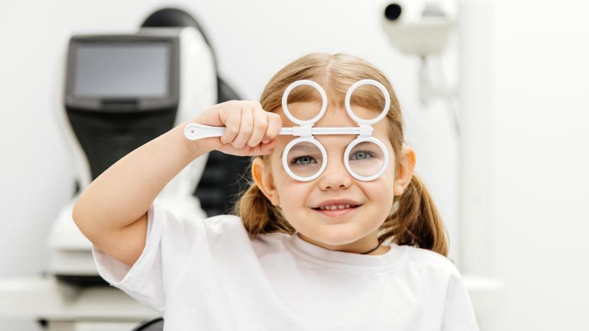
In this article, you’ll learn how to measure near lag of accommodation using practical, clinic-ready techniques. We outline the role of accommodative lag in the context of myopia, and how to measure near lag of accommodation using near retinoscopy as well as alternative techniques.
Role of accommodation in childhood myopia
Assessing accommodative function – including near lag of accommodation, is a vital component of understanding your patient’s risk profile for myopia. Accommodation and binocular vision status can influence myopia onset and progression in both children and adults.
Research suggests that esophoria and increased accommodative lag are associated with myopia compared to emmetropia.
As myopia development and progression is multifactorial, assessing accommodation and binocular vision contributes an important layer to clinical decision making.
How to measure accommodative lag with near retinoscopy
The simplest way to measure accommodative lag is with near retinoscopy, using the MEM retinoscopy technique. Watch this technique in action.
- Position yourself 33–40 cm from the patient, at their near working distance, to create an accommodative demand of 2.5–3 D.
- Ask the patient to look at your nose or at a near fixation card attached to your retinoscope. Ensure the patient is wearing full distance correction.
- Without a correcting lens in place, sweep the retinoscope horizontally and vertically in front of the pupil, and note the motion of the light reflex (e.g. with, against, or neutral).
- Repeat this step, but introduce lens flippers (±1.00, ±1.50 and ±2.00 flippers are useful) in order to neutralize the reflex.
- Try ±1.00 first, and if you still observe ‘with’ movement, move to ±1.50. If the reflex reverses, your answer is halfway between the two lenses such as +1.25.
- Identify the lens that produces neutralisation or the first reversal of movement – this final lens represents the accommodative response. No additional correction value is required.
Alternatively, Nott retinoscopy can be used, where the fixation plane stays constant and the examiner moves forwards or backwards to find the neutralised reflex.
Alternative ways to measure accommodative lag
Using a near fixation card
Some clinicians prefer to have the patient view a near fixation card as opposed to fixating on their nose, and this typically produces a slightly less plus result. There is no universally recommended method, so it may be practical to choose one approach (with or without a fixation card), and use it consistently to allow easy clinical comparisons.
Measuring lag with a fixation card can offer a picture that feels closer to real-world visual behaviour because it reflects active accommodation. On the other hand, measuring with a nose fixation target may reduce the influence of the test itself on subconscious accommodative tone.
Using fused cross-cylinder (FCC)
You can also measure accommodative lag with the fused cross-cylinder (FCC) card in the phoropter.
In practice, this method can sometimes produce results that are at odds with free-space techniques. Active accommodators may become aware of the phoropter in front of their face, which can influence the response and occasionally result in an accommodative lead on FCC, even when a lag has already been seen with near retinoscopy.
A previous study reported significantly lower accommodative response measurements with FCC compared to MEM or Nott retinoscopy, which aligns with these observations.
Using an open-field autorefractor
If you’re lucky enough to have an open-field autorefractor, you can measure accommodative lag using what is often considered the gold standard approach.
One study found good agreement between MEM and Nott retinoscopy, although both methods seemed to underestimate accommodative lag compared with open field autorefraction.
Final thoughts
Near retinoscopy remains a quick and practical way to measure accommodative lag. With a retinoscope and a set of flippers, you can typically obtain a reliable result in under 20 seconds, which gives you enough time to observe the stability of the reflex and neutralise it accurately.
Normal values fall between +0.50 and +1.00, with higher results indicating an increased risk for myopia.
Want to build your skills even further? Watch our video guides on how to assess accommodative facility and how to measure vergence in practice.
Key points
- Near retinoscopy, especially with the MEM method, provides a fast and reliable way to assess accommodative lag in routine clinical practice.
- Accommodative lag, and other binocular vision findings such as reduced facility and increased AC/A ratios can offer useful insight into a patient’s myopia risk profile.
- Alternative methods including near fixation cards, fused cross-cylinder, and open-field autorefraction can be used, although each technique has its caveats.
Want to learn more about binocular vision?
Discover our Binocular Vision Fundamentals online course – designed to build your confidence and clinical precision in assessing and managing BV.
You’ll learn our practical two-system approach to accommodation and vergence, step through key diagnostic tests, and refine your prescribing and management decisions. The course also explores how to communicate BV findings with patients and why BV is essential in myopia management – always with a sharp focus on clinical application.
Enjoy video demonstrations, real-world case examples, and downloadable chairside infographics you can use immediately in practice.
Meet the Authors:
About Kate Gifford
Dr Kate Gifford is an internationally renowned clinician-scientist optometrist and peer educator, and a Visiting Research Fellow at Queensland University of Technology, Brisbane, Australia. She holds a PhD in contact lens optics in myopia, four professional fellowships, over 100 peer reviewed and professional publications, and has presented almost 300 conference lectures around the world. Kate is the Chair of the Clinical Management Guidelines Committee of the International Myopia Institute. In 2016 Kate co-founded Myopia Profile with Dr Paul Gifford; the world-leading educational platform on childhood myopia management. After 13 years of clinical practice ownership, Kate now works full time on Myopia Profile.

About Brian Peng
Brian is a clinical optometrist based in Sydney, Australia. He graduated from the University of New South Wales and was awarded the Research Project Prize for his work on myopia. He has a keen interest in myopia-related research, industry, and education.
Read Brian's work on our My Kids Vision website, our public awareness platform. Brian also works on development of various new resources across MyopiaProfile.com.
References
- Drobe B, de Saint-André R. The pre-myopic syndrome. Ophthalmic Physiol Opt. Sep 1995;15(5):375-8. [link]
- Nakatsuka C, Hasebe S, Nonaka F, et al. Accommodative lag under habitual seeing conditions: comparison between myopic and emmetropic children. Jpn J Ophthalmol. Jan 2026;49(3):189-94. [link]
- Gwiazda J, Bauer J, Thorn F, et al. A dynamic relationship between myopia and blur-driven accommodation in school-aged children. Vision Res. May 1995;35(9):1299-304. [link]
- Allen PM, O'Leary DJ. Accommodation functions: co-dependency and relationship to refractive error. Vision Res. Feb 2006;46(4):491-505. [link]
- Harb EN, Thorn F, Troilo D. Characteristics of accommodative behavior during sustained reading in emmetropes and myopes. Vision Res. Aug 2006;46(16):2581-92. [link]
- Gwiazda J, Thorn F, Held R. Accommodation, accommodative convergence, and response AC/A ratios before and at the onset of myopia in children. Optom Vis Sci. Apr 2005;82(4):273-8. [link]
- Correction of Myopia Evaluation Trial 2 Study Group for the Pediatric Eye Disease Investigator Group , Manny RE, Chandler DL, et al. Accommodative lag by autorefraction and two dynamic retinoscopy methods. Optom Vis Sci. Mar 2009;86(3):233-43. [link]
- Locke LC, Somers W. A comparison study of dynamic retinoscopy techniques. Optom Vis Sci. Aug 1989;66(8):540-4. [link]
- Gwiazda JE, Hyman L, Norton TT, et al. Accommodation and related risk factors associated with myopia progression and their interaction with treatment in COMET children. Invest Ophthalmol Vis Sci. Jul 2004;45(7):2143-51. [link]
Enormous thanks to our visionary sponsors
Myopia Profile’s growth into a world leading platform has been made possible through the support of our visionary sponsors, who share our mission to improve children’s vision care worldwide. Click on their logos to learn about how these companies are innovating and developing resources with us to support you in managing your patients with myopia.